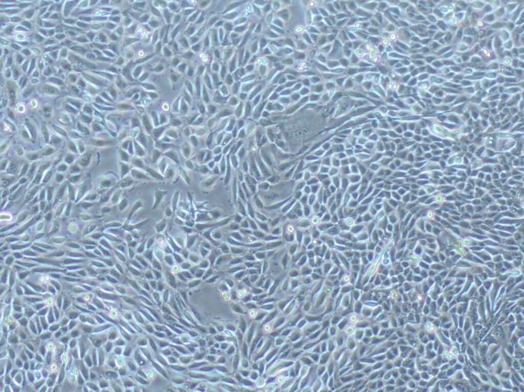
CLTH / PC, Primary Human Prostate Cancer Cell Line
WT, cin8Δ, and cin8-RAKA strains were analyzed for the percentage of cells in prometaphase and metaphase by microscopy (Figure 4E). There was a significant increase in cells delayed in mitosis in both cin8 mutant strains, consistent with a role for PP1 binding in mediating mitotic progression.
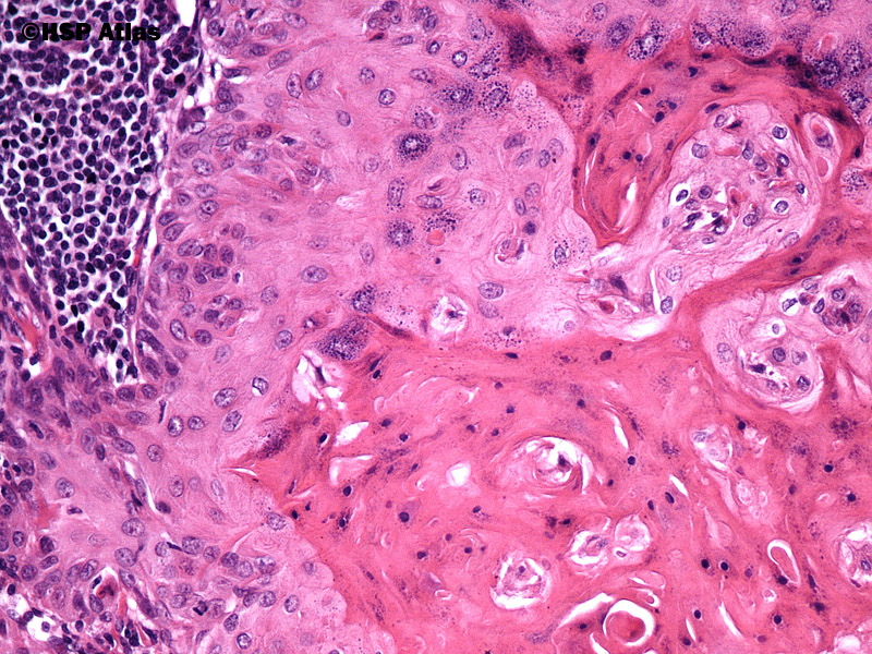
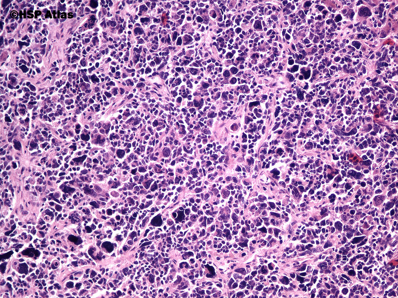
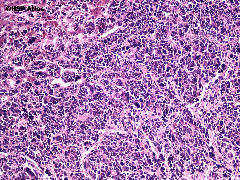
HSP Atlas Histopathology Atlas
RAK cells were transfused to primary HCC patients intravenously after RFA. During a seven-month follow-up, no severe adverse events, recurrences or deaths were observed, suggesting the feasibility and safety of the combined therapeutic strategy to prevent HCC recurrence . In 2008, Weng et al. launched a clinical trial which recruited 85 HCC.
Rak może “zapadać w sen”, żeby ochronić się przed chemioterapią
People named Raka Cell. Find your friends on Facebook. Log in or sign up for Facebook to connect with friends, family and people you know. Log In. or. Sign Up. Raka Riki Cell. See Photos. Raka Cell. See Photos. @raka.embarkasi. Sukoharjo, Jawa Tengah, Indonesia. Rakan Cell. See Photos.
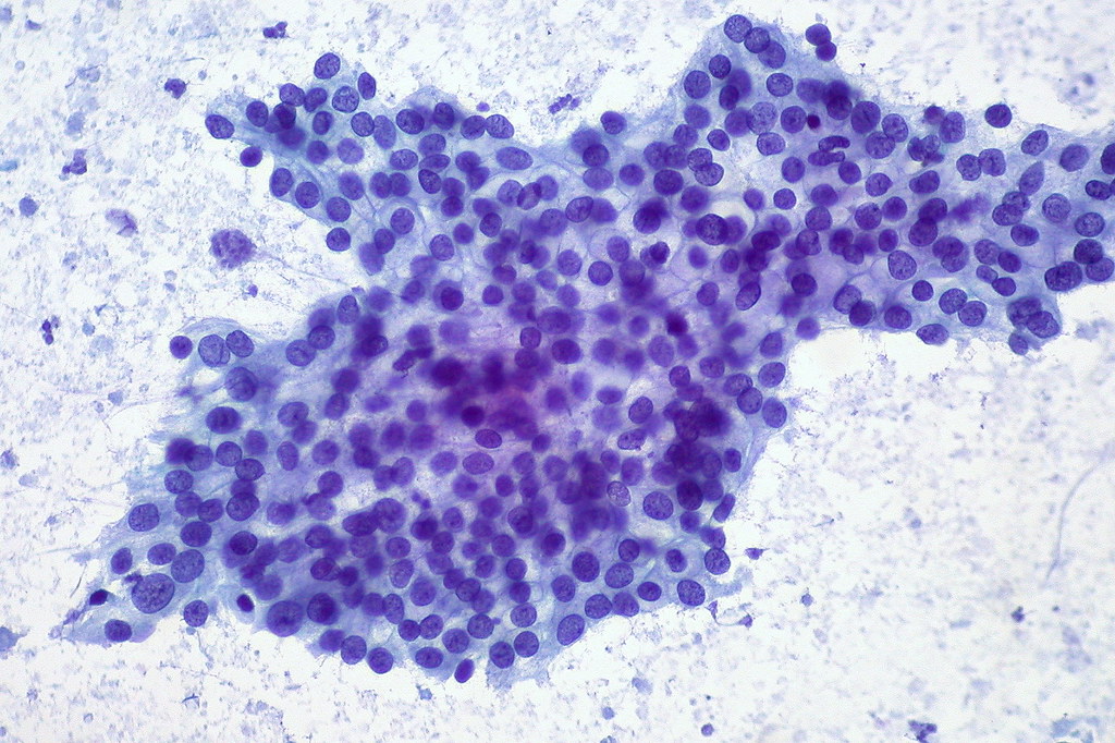
Adenocarcinoma of Pancreas, FNA This is one of the better … Flickr
Rak Suppresses Tumorigenicity of Human Breast Cancer Cells. Since we found that Rak regulates the stability of PTEN protein and that mutation of the Rak phosphorylation site on PTEN decreases the growth-inhibitory effect of the PTEN tumor suppressor gene, we posited that Rak might function as a tumor suppressor gene.
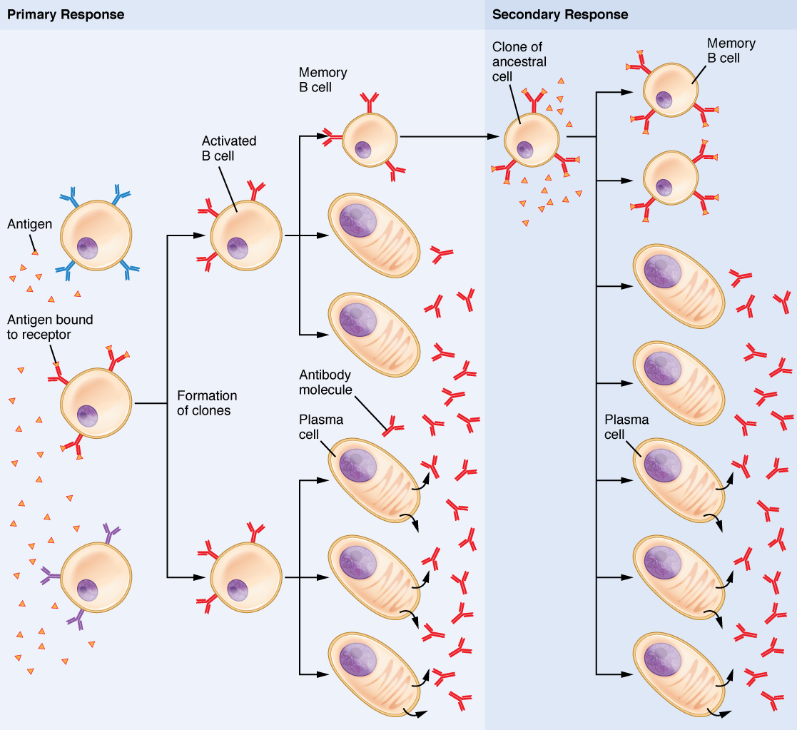
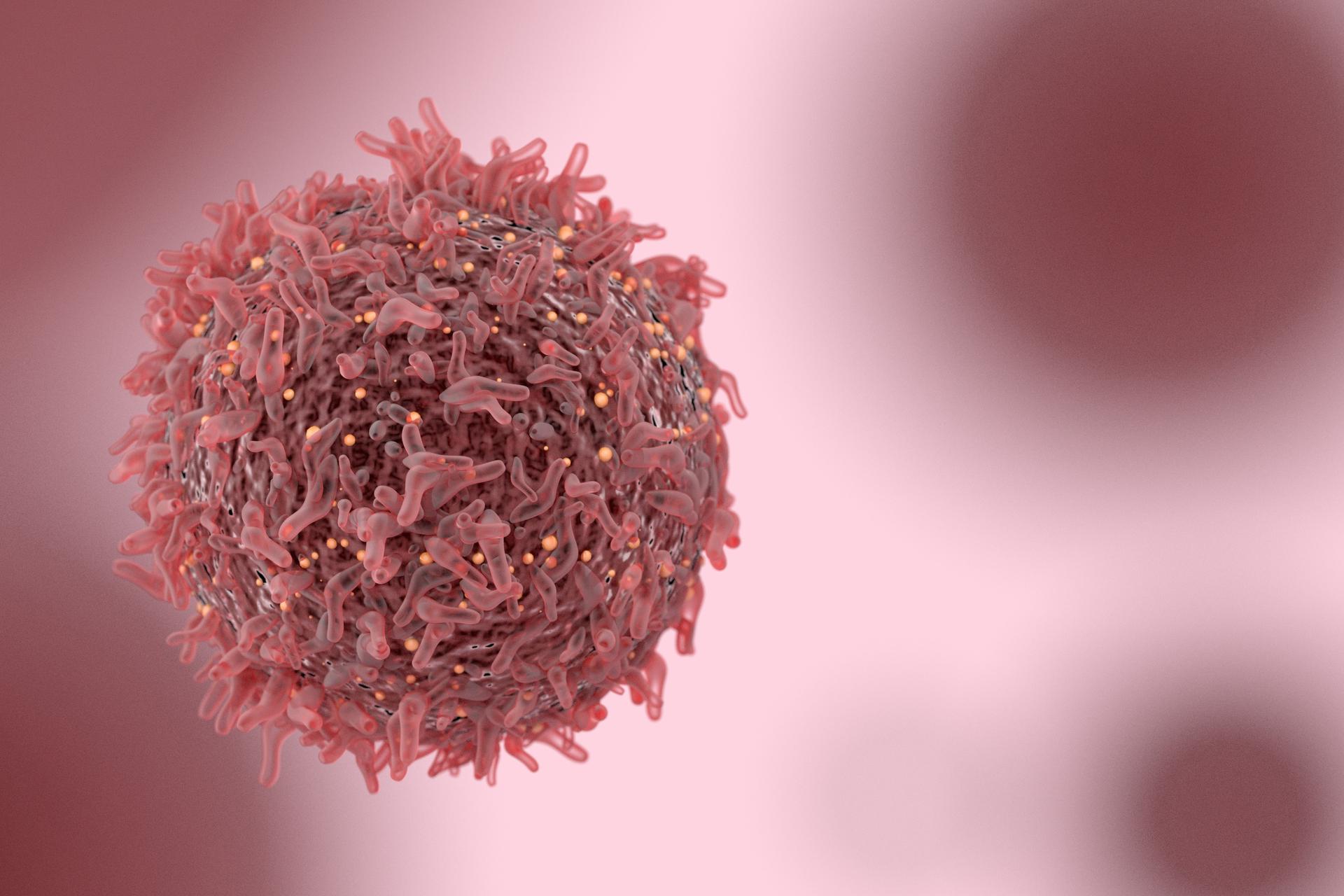
The Adaptive Immune Response Blymphocytes and Antibodies · Anatomy
We hypothesis pazopanib plus PD-1 inhibitor and RAK cells together could bring more benefit in the treatment of our patient. RAK cells is a new kind of cytokine induced killer cells which use the proliferation technique of RetroNectin [17, 18]. One study found that RAK cells are heterogeneous cells in CD3 + CD8 + CD56 + T cells. These cells.
Yale Cancer Center researchers show mutations in advanced lung
Non-small cell lung cancer (NSCLC) is a frequent type of cancer, which is mainly characterized clinically by high aggressiveness and high mortality. KRAS oncoprotein is the most common molecular protein detected in NSCLC, accounting for 25% of all oncogenic mutations. Constitutive activation of the KRAS oncoprotein triggers an intracellular cascade in cancer cells, leading to uncontrolled cell.
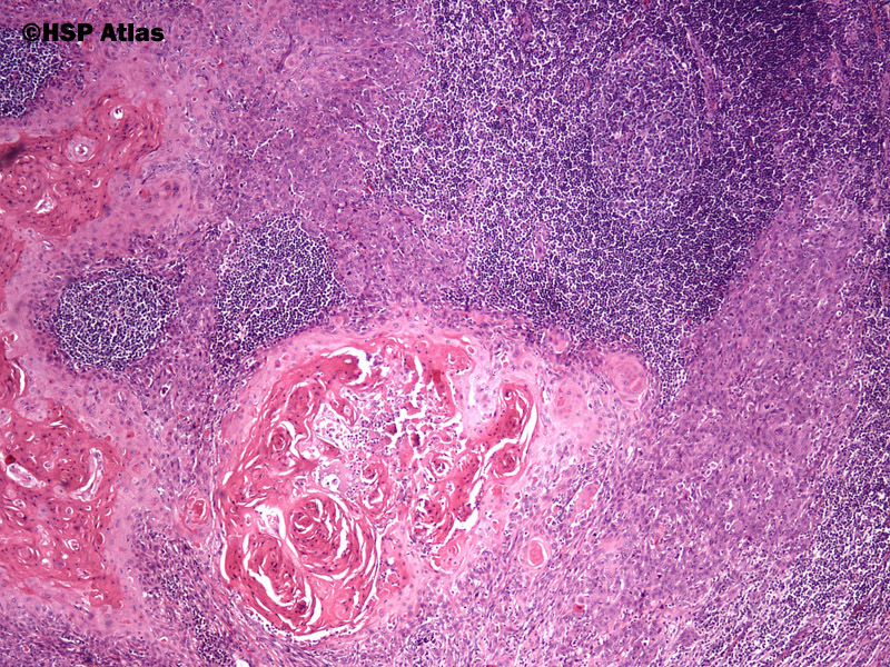
HSP Atlas Histopathology Atlas
On (±2) days 15 and 16, Autologous RAK cells were reinfused for treatment (cycle 1). TACE was performed on day 28 (±7). Blood drawing from autologous RAK cells in the second cycle (week 10 ±7 days) The second cycle of autologous RAK cell transfusion treatment (12 weeks ±7 days after hepatocellular carcinoma surgery); Blood was drawn from.
Rano otkivanje raka debelog creva ključno u borbi protiv ove bolesti
Rak in breast cancer cells. Rak Regulates PTEN Protein Stability Next, we sought to determine whether there is a causal relation-ship between Rak status and PTEN protein levels. To this end, we established two Rak-overexpressing MCF7 breast cancer cell lines (Rak 44 and Rak 45; Figure 1F, left) and three stable
Nowotwór czy rak? Jak to jest czy każdy nowotwór to rak
An inflammatory immune cell model was constructed by exposing RAW 264.7 cells to LPS. A series of experiments, including biochemical assays, RT‒PCR, and western blotting, were conducted to investigate the anti-inflammatory and antioxidative effects of PREO.. Rifat Nowshin Raka, Ding Zhiqian & Xiao Junsong. College of Chemical and Materials.
HSP Atlas Histopathology Atlas GI Tract
Background: Primary hepatic angiosarcoma (PHA) is a rare and aggressive solid tumor, with high rates of local recurrence and distant metastasis, and poor prognosis. There are no established treatment guidelines for PHA. Case presentation: A 78-year-old asymptomatic man with PHA that was successfully treated with pazopanib plus PD-1 inhibitor and RetroNectin-activated killer cells (RAK cells).
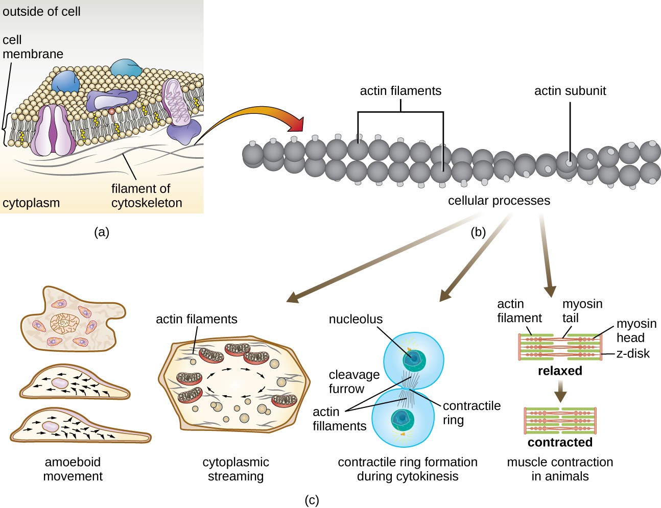
Unique Characteristics of Eukaryotic Cells Microbiology
As shown in Figure Figure1A, 1A, Rak-depleted MCF10A cells (siRak#1 and #3) exhibited an increase in the number of γH2AX foci compared to control cells, suggesting that Rak deficiency causes an accumulation of spontaneous DNA DSBs. The induction of DSBs in the absence of Rak was also confirmed by the neutral comet assay.
HSP Atlas Histopathology Atlas Urinary
Decades of oncologic clinical use have demonstrated that cancer immunotherapy provides unprecedented therapeutic benefits. Tragically, only a minority of patients respond to existing immunotherapies. RNA lipid nanoparticles have recently emerged as modular tools for immune stimulation. Here, we discuss advancements in RNA-based cancer immunotherapies and opportunities for improvement.
Test na raka WP Tech
Quality of RAK cells. The final cell products were assessed for viability by the dye-exclusion test and checked twice for possible contamination by bacteria, fungi and endotoxins. The propor-tions of viable RAK cells exceeded 95%. No contamination was found. Before RAK cell administration, lymphocyte subsets were measured.
Przerzuty raka powstrzymywane przez lepkie nanocząsteczki Artykuły
The FRET emission ratio at metaphase was significantly higher (∼3.3) in cin8-RAKA cells than in the control (∼2.2), indicating that an almost complete loss of tension as occurs with cin8Δ cells and Ndc80 tailless mutants (Figures 3 B and 4 F) [4]. We note that cin8-RAKA cells did not alter the level of Cin8 at kinetochores (Figure S3 C).
Immunoterapia raka kory nadnercza badanie kliniczne
Control or Rak-knockdown cells were seeded in a 96-well plate at 1 × 10 4 cells per well. Cell proliferation was measured by MTT assay for 5 days. (B) Rak knockdown induces anchorage-independent growth of MCF10A cells. Viable colonies of MCF10A clones in three wells were counted; all soft agar assays were performed in triplicate..
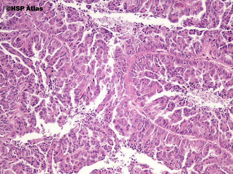
HSP Atlas Histopathology Atlas GI Tract
Washed, stained cells were mounted using Vectastain mounting media (Vector Labs) and fluorescence-visualized through the GFP filter on a Leica DM5000B microscope and quantified using ImageJ. Conditioned Media Experiments. Primary CAPSCs were grown to 100% confluency. Fresh media was added to the cultures, and at this time, CAPSCs were treated.
